| dc.contributor.author | Özbek, Zühtü | |
| dc.contributor.author | Koçman, Atacan Emre | |
| dc.contributor.author | Özatık, Orhan | |
| dc.contributor.author | Söztutar, Erdem | |
| dc.contributor.author | Özkara, Emre | |
| dc.contributor.author | Köse, Aydan | |
| dc.contributor.author | Arslantas, Ali | |
| dc.contributor.author | Çetin, Cengiz | |
| dc.date.accessioned | 12.07.201910:49:13 | |
| dc.date.accessioned | 2019-07-11T21:57:59Z | |
| dc.date.available | 12.07.201910:49:13 | |
| dc.date.available | 2019-07-11T21:57:59Z | |
| dc.date.issued | 2017 | |
| dc.identifier.issn | 1019-5149 | |
| dc.identifier.uri | https://app.trdizin.gov.tr/makale/TWpNd056UXdNQT09 | |
| dc.identifier.uri | https://hdl.handle.net/20.500.12513/1149 | |
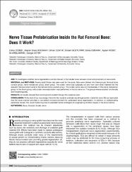
| dc.description.abstract | AIm: To investigate whether nerve regeneration can be induced in the tubular bone between distal and proximal cut nerve ends. mATERIAl and mEThODS: Twenty adult Wistar rats were used for the study. Rats were divided into three groups; femoral bone conduit group, nerve transection group, sham group. The sciatic nerve was surgically cut and from both ends inserted into the adjacent femoral bone tunnel in the femoral bone conduit group. The sciatic nerve was cut transversely in the nerve transection group. In the Sham group, only sciatic nerve exploration was performed, without a nerve cut. The groups were evaluated functionally and morphologically. RESUlTS: All results showed that axonal growth existed through the osseous canal. CONClUSION: To the best of our knowledge, this is the first study to evaluate neural regeneration inside the bone. We can speculate that the bone marrow provides a convenient microenvironment for peripheral nerve regeneration. In addition to prefabricating peripheral nerves, this novel model may help to establish further strategies for engineering of other tissues in the bone marrow | en_US |
| dc.description.abstract | AIm: To investigate whether nerve regeneration can be induced in the tubular bone between distal and proximal cut nerve ends. mATERIAl and mEThODS: Twenty adult Wistar rats were used for the study. Rats were divided into three groups; femoral bone conduit group, nerve transection group, sham group. The sciatic nerve was surgically cut and from both ends inserted into the adjacent femoral bone tunnel in the femoral bone conduit group. The sciatic nerve was cut transversely in the nerve transection group. In the Sham group, only sciatic nerve exploration was performed, without a nerve cut. The groups were evaluated functionally and morphologically. RESUlTS: All results showed that axonal growth existed through the osseous canal. CONClUSION: To the best of our knowledge, this is the first study to evaluate neural regeneration inside the bone. We can speculate that the bone marrow provides a convenient microenvironment for peripheral nerve regeneration. In addition to prefabricating peripheral nerves, this novel model may help to establish further strategies for engineering of other tissues in the bone marrow | en_US |
| dc.language.iso | eng | en_US |
| dc.rights | info:eu-repo/semantics/openAccess | en_US |
| dc.subject | Cerrahi | en_US |
| dc.title | Nerve Tissue Prefabrication Inside the Rat Femoral Bone: Does It Work? | en_US |
| dc.type | article | en_US |
| dc.relation.journal | Turkish Neurosurgery | en_US |
| dc.contributor.department | Kırşehir Ahi Evran Üniversitesi | en_US |
| dc.identifier.volume | 27 | en_US |
| dc.identifier.issue | 4 | en_US |
| dc.identifier.startpage | 648 | en_US |
| dc.identifier.endpage | 655 | en_US |
| dc.relation.publicationcategory | Makale - Ulusal Hakemli Dergi - Kurum Öğretim Elemanı | en_US] |